Describe the Wall of the Urinary Bladder of the Cat
Cats with urinary obstruction may strain hard enough to increase bladder pressure above the breaking strength of the bladder wall and cause a rupture. No acoustic shadowing is.

Front Line Ultrasound Imaging Of The Feline Urinary
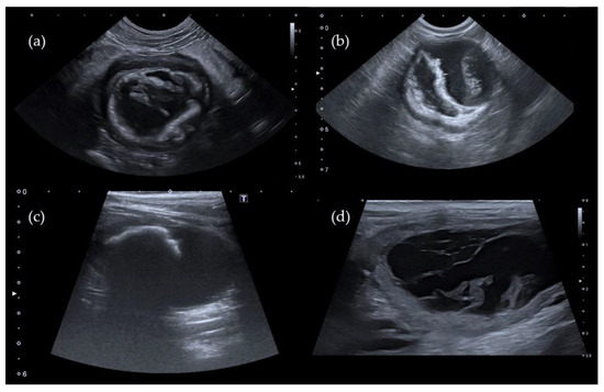
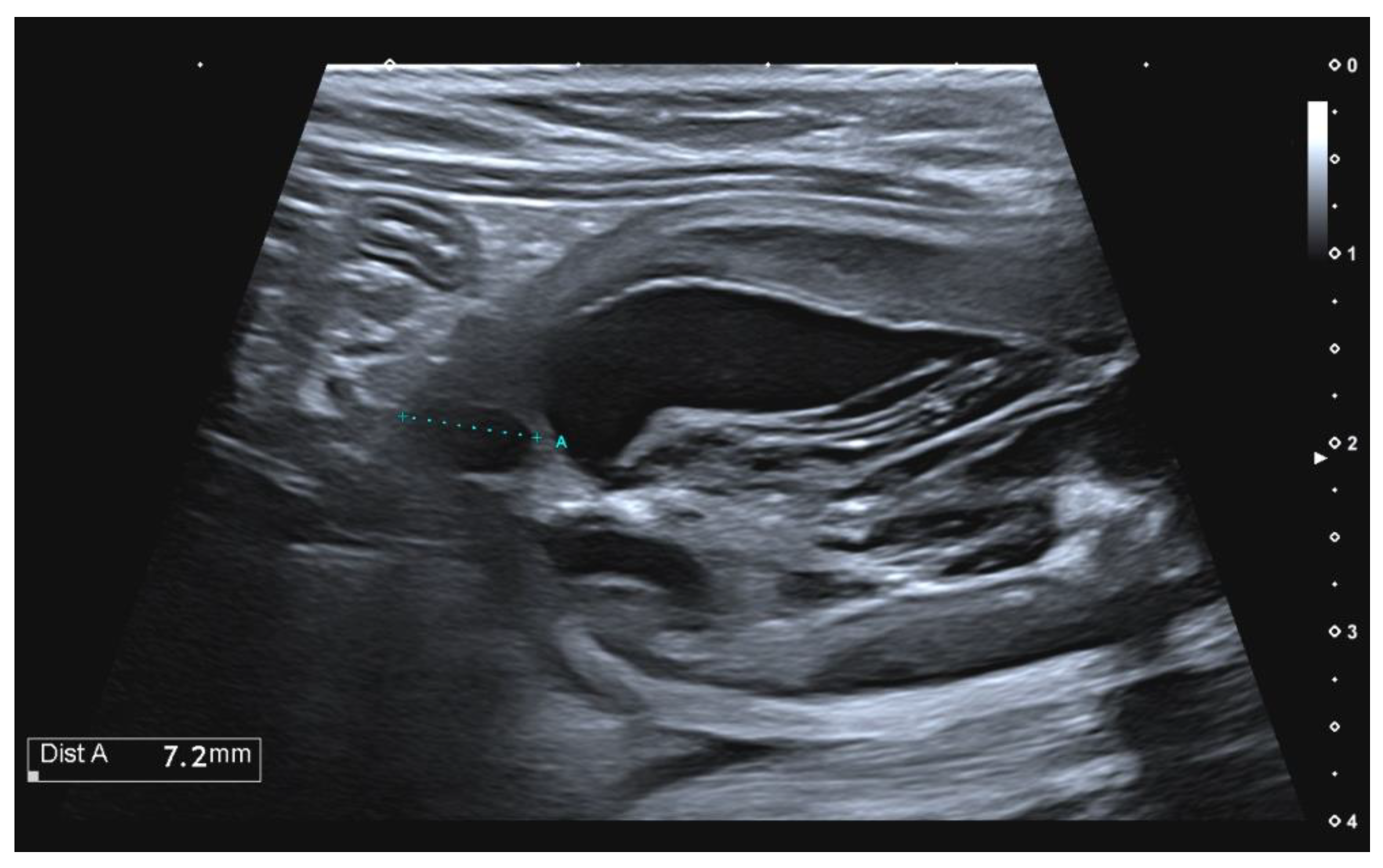
Note the dependently located hyperechoic grass awn in the lumen white arrows.
. Include the thickness texture and inner surface in youranswer. Compare the renal pyramids and renal papillae of the human kidney with those of the cat. The cats urinary tract is a system made up of the kidneys ureters urinary bladder and urethra.
Ultrasound shows that the blood clot is quite echogenic unlike a bladder tumor which often has an echogenicity similar to the bladder wall or other soft tissue. In previous reports lymphoma affecting the urinary bladder was localized in the ventral urinary bladder wall in a dog 7 in the dorsocranial aspect of the bladder wall in a cat 8 and at the. Mucous membrane - Urothelium prev.
24921 In this cat abdominal changes were an. Muscular wall is expandable and contractible. Peristalitic waves convey urine along the tube.
Transitional epithelium-Apical cellular protection against urine-Wide range of adaptation if wall is stretched Lamina propria Muscular layer Adventitia. Contrast radiography is often necessary to evaluate the lower urinary tract. List the components of the nephron from proximal to distal.
The musculomembranous urinary bladder wall consists of mucosal submucosal and muscular layers. A triangular area of the floor of the urinary bladder between the opening of the urethra in front and the two ureters at the sides. B The trigone and the urethra appear normal Capasso et al 3.
Compare the locations of the adrenal glands in the cat with those in the human. The thickness of the bladder wall or mucosa cannot be identified on survey radiographs because of border effacement by urine in the lumen. However on plain films a thickened bladder wall a distended bladder or a displaced urinary bladder and renal changes can be observed.
Mucosa submucosa muscularis and serosa or adventitia. Wall made up from. Science Biology QA Library Describe the wall of the urinary bladder of the fetal pig.
For a healthy continent cat a full bladder is somewhere between the size of a handball and a golf ball while the bladder of an incontinent cat can sometimes be as large as a large grapefruit. Ureter Locate the large bean shaped kidney along the dorsal surface of the abdominal cavity. Some incontinent cats can have larger bladders because over time the bladder walls have stretched to accommodate the retained urine.
Some authorities do not include a submucosa in the wall of the urinary bladder--in their opinions all of the connective. The urinary bladder is found inferior to the peritoneum sitting on the pelvic floor. Locate the following urinary structures and describe their functions.
Muscle that forms the layer of the wall of the bladder. A1 4 Describe the wall of the urinary bladder ofthe cat. The wall of the urinary bladder has four layers.
Sagittal image of the urinary bladder of Cat 1. In males the inferior surface of the bladder lays over the pubic symphysis and prostate posteriorly is the distal third of the rectumBetween the posterior surface of the. Describe the wall of the urinary bladder of the fetal pig.
However on plain films a thickened bladder wall a distended bladder or a displaced urinary bladder and renal changes can be observed. The peritoneum is adherent to the serosal surface providing a separate fourth layer. Border of kidney that is toward the midline medial has hilus.
Include the thickness texture and inner surface in youranswer. 24921 In this cat abdominal changes were an. Clots in the bladder may have a vermiform or wormlike appearance if bleeding has occurred in the upper urinary tract and a clot has formed in the ureter.
These organs work together to produce transport store and excrete urine. 2 bean shaped organs located toward the dorsal body wall surface and behind peritoneum. Remove nitrogenous waste from blood balance body fluids and form urine.
Obstruction can occur secondary to stones that form within the bladder and get lodged in the urethra or due to masses growing within the pelvic canal that compress the urethra itself. A An irregular rounded 46 42 4 cm lesion arises from the apex and the dorsal wall of the urinary bladder. In cats with urinary bladder tumours abdominal radiographs are mainly unremarkable.
Contrast radiography is often necessary to evaluate the lower urinary tract. From the inside towards the outside they are. What structures of the cat urinary system are retroperitoneal.
In females its inferior surface lays on the pubic symphysis and the posterior wall is in contact with the vagina and uterus. Muscular tube that conveys urine form pelvis of the kidney to urinary bladder 2. The urinary tract also rids the body of many fluid waste materials and products and has other vitally important functions including controlling the volume and composition of the body fluids.
The inner wall of the bladder is called urothelium a type of transitional epithelium formed by three to six layers of cells. Carefully cut away the surrounding fat and trace the ureter to the bladder. The outside layer is either serosa or adventitia depending on location--see your textbook for an explanation.
6 9 1011 Clinical. Border of kidney that is away from the midline lateral medial border. In cats with urinary bladder tumours abdominal radiographs are mainly unremarkable.
A primary fibrosarcoma of the urinary bladder in a cat has been recently described and it has been hypothesised that it might have a different behaviour than in dogs. Now remove one kidney from the cat. The cells may become more cuboidal or flatter depending on whether the bladder is empty or full.
The Urinary System Of Cats Cat Owners Msd Veterinary Manual

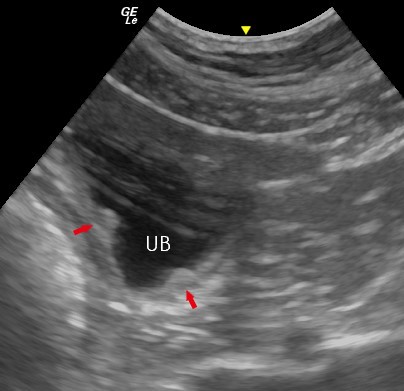
Sagittal Image Of The Urinary Bladder Of Dog 1 Note The Dependently Download Scientific Diagram

Small Animal Abdominal Ultrasonography The Urinary Tract Urinary Bladder And Urethra Today S Veterinary Practice
The Urinary System Of Cats Cat Owners Msd Veterinary Manual

Small Animal Abdominal Ultrasonography The Urinary Tract Urinary Bladder And Urethra Today S Veterinary Practice
2
Front Line Ultrasound Imaging Of The Feline Urinary

Small Animal Abdominal Ultrasonography The Urinary Tract Urinary Bladder And Urethra Today S Veterinary Practice
![]()
Sagittal Image Of The Urinary Bladder Of Cat 1 Note The Dependently Download Scientific Diagram
Veterinary Sciences Free Full Text Pseudomembranous Cystitis An Uncommon Ultrasound Appearance Of Cystitis In Cats And Dogs Html
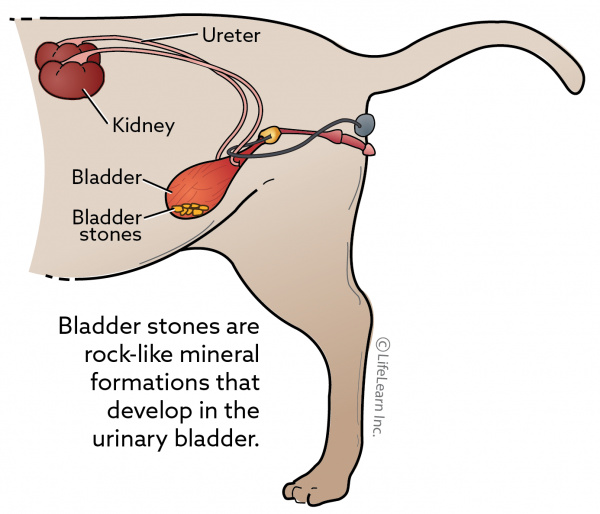
Bladder Stones In Cats Vca Animal Hospital

Fibrosarcoma Of The Urinary Bladder In A Cat Semantic Scholar

Urinary Health Of Cats Evolution Diet
Ultrasonography Of The Urinary Bladder Imv Imaging

Primary Fibrosarcoma Of The Urinary Bladder In A Cat Follow Up After Incomplete Surgical Excision
Veterinary Sciences Free Full Text Pseudomembranous Cystitis An Uncommon Ultrasound Appearance Of Cystitis In Cats And Dogs Html

Small Animal Abdominal Ultrasonography The Urinary Tract Urinary Bladder And Urethra Today S Veterinary Practice

Sagittal Image Of The Urinary Bladder Of Cat 1 Note The Dependently Download Scientific Diagram

Solved Laboratory Name Assessment Date Sectionn 67 Cat Chegg Com


Comments
Post a Comment